The renal arteries are instrumental in supplying blood to the kidneys. The occurrence of renal artery disease, specifically the constricting or narrowing known as stenosis, generally instigated by atherosclerosis, can contribute to a diminished blood supply to the kidney. This condition can prompt the onset of hypertension, or high blood pressure. In fact, renal artery stenosis is frequently the most amendable cause of hypertension. Prolonged, untreated renal artery disease is also a key culprit in kidney failure.
Identifying renal artery disease necessitates specific tests, among which a renal artery duplex scan forms an accurate, non-invasive, and economical choice. Outperforming alternatives like angiography or CT scanning, the renal artery duplex scan requires no injection of X-ray contrast material, thus mitigating potential kidney damage from the contrast.
Renal Artery Duplex Scan: Causes And Risk Factors
The renal artery duplex scan is an ultrasound-based diagnostic test that explores potential issues within the renal arteries—blood vessels responsible for supplying your kidneys. By utilizing ultrasound imaging, the procedure portrays vivid images of the kidneys and arteries. A specialized Doppler technique integrated with the procedure measures blood movement within the vessels, offering crucial insights into any renal artery narrowing and its extent as well as cause.
Renal artery disorders tend to mask their presence until an advanced stage. Generally, their presence surfaces through symptoms like escalated blood pressure since the kidneys are responsible for regulating blood flow through the renal artery and thus, adjusting the body’s blood pressure.


Several underlying causes may lead to renal artery constriction or stenosis.
A common culprit is atherosclerosis, a plaque buildup caused by factors like aging, hypertension, and dyslipidemia (cholesterol abnormalities). This condition leads to progressive arterial narrowing and can occasionally result in sudden spikes in blood pressure. By employing a renal artery duplex scan, the presence of this disorder can be detected, thereby aiding interventional radiologists in deciding the best course of treatment, such as a renal artery stent implant.
Fibromuscular Dysplasia (FMD), another possible cause, leads to abnormalities in the renal arteries’ walls, which can trigger renal artery narrowing (stenosis) or enlargement (aneurysm). It can further impact other arteries in your body, potentially reducing organ function due to diminished blood flow. Predominantly observed in the arteries leading to the brain and kidneys, FMD may present as renal artery blockage. Interventional radiologists employ the renal artery duplex scan as an initial screening tool and use the findings to lead FMD treatment, like addressing any aneurysm or stenosis.
The onset of sudden hypertension (high blood pressure) before the age of 30 or after 50, or difficult-to-manage hypertension could indicate the need for treatment for renal stenosis. The notable risk factors encompass:
- Aging
- Diabetes
- Early heart disease in family history
- High blood pressure
- High cholesterol
- Limited physical activity
- Obesity
- Smoking or consumption of other tobacco products
- Renal Artery Duplex
- Vascular Disease Examination
- Ankle Brachial Index
- Aortic Ultrasound
- Aortoiliac Duplex
- CT Angiogram
- Renal Artery Duplex
- Venous Duplex
- Trinity Health Michigan’s proficient medics conduct the renal artery duplex scan as a preferred diagnostic approach to pinpoint any issues present in the kidney’s veins and arteries.
Explaining Renal Artery Duplex
The renal artery duplex scan is a non-invasive diagnostic exam harnessing ultrasound technology to peep into the kidney’s veins and arteries, scouting for potential narrowing or occlusions. Typically, this procedure runs between 60 to 90 minutes.
Renal artery disease is the most prevalent cause of the constriction of renal arteries. Unattended, diminished blood flow to the kidney can result in hypertension and potentially, kidney failure. If diagnosed via the renal artery duplex scan, your attending physician will work alongside you to map out a tailor-made treatment strategy, which could involve minimally invasive interventions or even surgery.
Noticeable symptoms for renal artery disease are usually absent, making it crucial to routinely monitor blood pressure to detect any abrupt escalations.
Understanding the Renal Artery Duplex Scan:
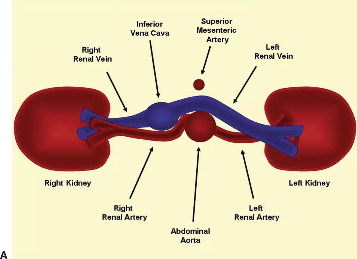
A renal artery duplex employs high-frequency sound waves to generate cross-sectional images of the arteries supplying the kidneys, encompassing the abdominal Aorta. The examination incorporates Doppler technology, a subcategory of ultrasound imaging, to assess blood flow within these arteries and the Aorta. The fusion of these imaging methods provides physicians with an in-depth look at the arterial condition, assisting them in detecting plaque, aneurysms, and potential blockages. The renal arteries are vital as they deliver blood to the kidneys. The Aorta, based within the abdomen, ensures the other abdominal organs receive an adequate blood supply.
Procedure of the Renal Artery Duplex Scan:
As part of the procedure, you will be requested to partially disrobe and recline on an exam table. A non-greasy gel will be spread over your abdomen, and then the sonographer will carefully maneuver an ultrasound probe across it, changing its direction as needed. Breath-holding is often involved, and you may also be asked to lie on your sides. Throughout the examination, you might hear distinct whip-like or whooshing sounds, which are characteristic of the Doppler phase of the scan. The renal artery duplex scan carries no pain; hence if any discomfort arises, alert the sonographer promptly to make necessary adjustments.
Post-Scan Process:
Upon the renal artery duplex scan’s completion, the captured images and measurements are forwarded to our skilled vascular cardiologist for evaluation. A comprehensive report, embodying the results and any suggestions for additional treatment, will be compiled and communicated to you. This report will also be integrated into your electronic Virginia Heart chart, accessible to your cardiologist for review. The document will likewise be dispatched to your referrer’s Physician. If you wish for any other healthcare professionals to receive your scan results, please inform the receptionist to make arrangements for a release that would permit distribution of your report.
Purpose of the Renal Artery Duplex Scan:
The main goal of the renal artery duplex is to utilize ultrasound technology to examine the arteries deriving from the abdominal aorta that supply blood to the kidneys. These arteries risk becoming abnormally narrowed (stenosis) or dilated (aneurysm). Stenosis may result in deteriorating renal function or elevated blood pressure, while aneurysms may rupture and bleed if left untreated. The duplex ultrasound uses sound waves to assess the artery’s morphology and scrutinize the blood flow within the artery, hence delivering two kinds of data, hence the term ‘Duplex’. This information aids your doctor in diagnosis and treatment planning, avoiding the need for needle-based or invasive procedures. Bear in mind the renal artery duplex scan is merely a diagnostic tool and does not directly treat arterial anomalies.
Preparation for the Renal Artery Duplex Scan:
Ensuring an accurate renal artery duplex necessitates that the patient fasts for a period of 6 to 8 hours prior to the scan. This is because ingested food and gastrointestinal gases may disrupt the technician’s visualization of the kidneys, which are deeply located in the abdomen, adjacent to the spine.
The Renal Artery Duplex Scan Technique:
For the renal artery duplex, the patient will lie on an examination bed with only the abdomen exposed. The technician will apply ultrasound gel and employ an ultrasound probe on the abdominal wall, adjusting the angle as needed to steer the sound waves in various directions. Reflected sound waves bouncing off diverse abdominal organs are captured by the probe, and these waves are converted into images on the computer monitor for analysis.
Time Duration:
The renal artery duplex scan typically lasts for approximately 60 minutes.
Diagnostic Critique:
From a diagnostic perspective, a stenosis exceeding 60% is inferred if the ratio of the peak velocity within the narrowed renal artery relative to the velocity within the aorta is above 3.5.
Also read: Sinus rhythm with artifact